- The paper introduces a novel confocal imaging system integrated in a dilution refrigerator to image nanophotonic devices at sub-Kelvin temperatures.
- It employs an 8f configuration with non-magnetic optics, achieving 1.1 μm resolution and a 2.5 mm field of view while compensating for thermal contractions.
- The methodology supports real-time imaging for quantum network node development, facilitating in-situ device characterization and fiber coupling.
Introduction
This work presents a robust confocal imaging system integrated into a dilution refrigerator, designed for high-resolution visualization of nanophotonic structures on transparent diamond substrates at sub-Kelvin temperatures. The system addresses the limitations of conventional cryogenic microscopy for quantum technology applications, where stringent control over temperature, vacuum, and magnetic fields is required. The imaging setup achieves a resolution of 1.1μm and a field-of-view (FOV) of $2.5$ mm, with no moving parts at cryogenic temperatures and a large working distance to facilitate probe access and sample anchoring. This configuration is particularly relevant for the development and characterization of quantum network nodes and optically connected quantum processors.
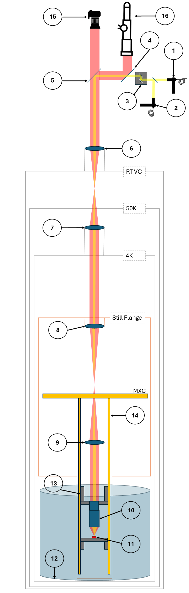
Figure 1: Schematic of the dilution refrigerator with all imaging components, illustrating the optical path and integration of the imaging system.
System Architecture and Design Considerations
The imaging system is based on an $8f$ confocal configuration, utilizing a commercial 10× infinity-corrected objective lens positioned within the bore of a superconducting vector magnet at the mixing chamber stage. Four relay achromatic lenses form two $4f$ subsystems, transferring the collected light across the cryostat's temperature stages. The final relay lens also serves as the vacuum window, ensuring both optical transmission and thermal isolation. External tube lenses and motorized focusing mechanisms allow for post-cooldown focal adjustment, compensating for thermal and pressure-induced shifts.
Key design choices include:
- Non-magnetic, high-resolution objective: Avoids magnetic interference and enables compatibility with high-field environments.
- Fixed optics at cryogenic temperatures: Eliminates vibration-induced misalignment and ensures stable imaging under ultra-high vacuum.
- Confocal illumination via galvanometer scanning: Enhances contrast for low-visibility nanophotonic structures and enables hyperspectral imaging.
- Large working distance: Facilitates integration of fiber-optic and microwave probes for device characterization and coupling.
Compensation of Focal Shifts and System Response
Thermal contraction, changes in refractive index, and pressure variations during evacuation and cooldown induce focal plane shifts. The system compensates for these effects by pre-adjusting the objective position at ambient conditions and utilizing motorized tube lenses for post-cooldown correction. The focal shift Δstel is related to the sample-objective distance shift Δsobj by Δstel≈−M2Δsobj, where M is the magnification.
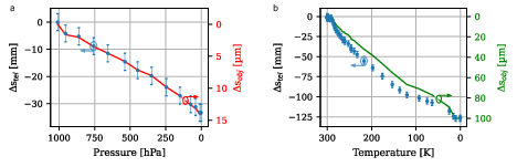
Figure 2: Change of focal plane due to pressure (a) and temperature (b) variations, showing measured and extrapolated shifts during evacuation and cooldown.
Experimental characterization reveals that the observed focal shift during cooldown (∼0.10 mm) is within the correctable range of the tube lens system. The shift is approximately linear in temperature, attributed primarily to thermal contraction, with minor contributions from refractive index changes and pressure effects. The system demonstrates repeatable compensation across thermal cycles, validating the robustness of the pre-compensation strategy.
Resolution was quantified using USAF-1951 test targets and Modulation Transfer Function (MTF) analysis. At room temperature, the system achieves ∼800 lp/mm, while at cryogenic temperatures (<100 mK), resolution is reduced to ∼400 lp/mm, corresponding to 1.1μm. The reduction is attributed to aberrations from lens position shifts and off-axis astigmatism, which can be partially mitigated by careful alignment and objective tilt.
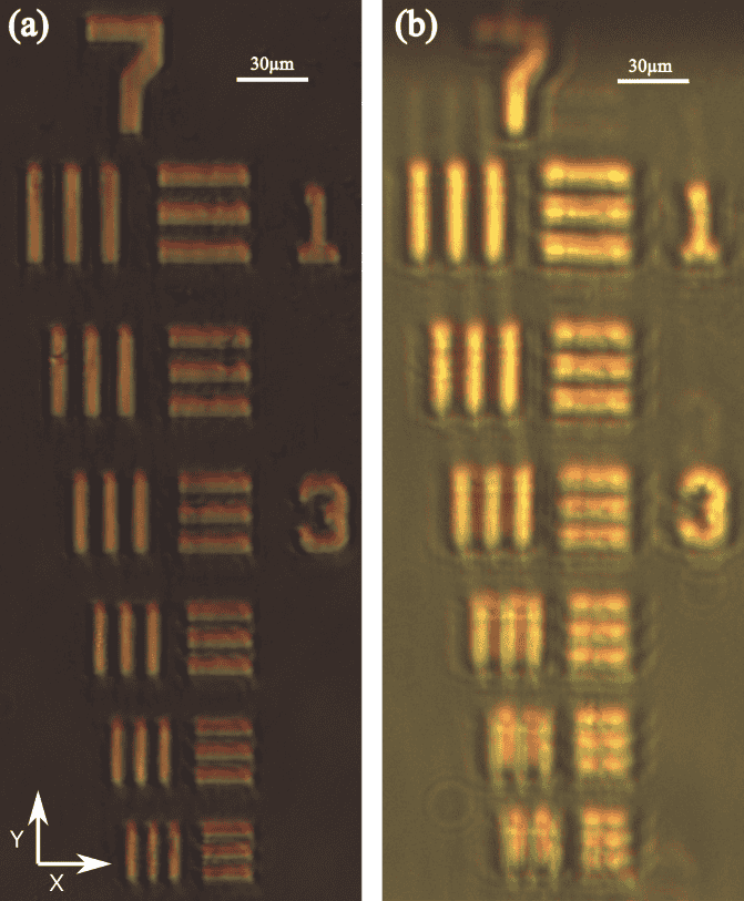
Figure 3: Exemplary images of the USAF-1951 test target at room temperature (a) and cryogenic temperature (b), demonstrating maintained resolution.
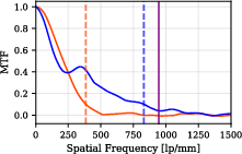
Figure 4: MTF analysis for the Y-axis at ambient and cryogenic conditions, indicating the resolution drop and technical limits.
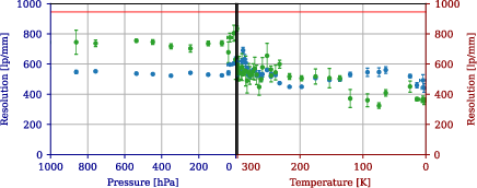
Figure 5: Change of resolution during evacuation and cooldown, with error bars representing statistical variance from repeated optimization.
Astigmatism and field curvature are more pronounced for off-axis regions, especially given the non-plane-corrected objective and sample tilt. The wide-field imaging system provides rapid FOV adjustment, while the high-magnification setup enables detailed inspection of nanophotonic features.
Application to Nanophotonic Device Characterization
The imaging system was employed to visualize nanophotonic structures on diamond chips at ∼10 mK. Waveguides ($550$ nm wide, 125μm long) and alignment markers are clearly resolved, and supports as narrow as 1.0μm are discernible. The system facilitates real-time monitoring of fiber coupling procedures and device alignment, critical for quantum interface development.
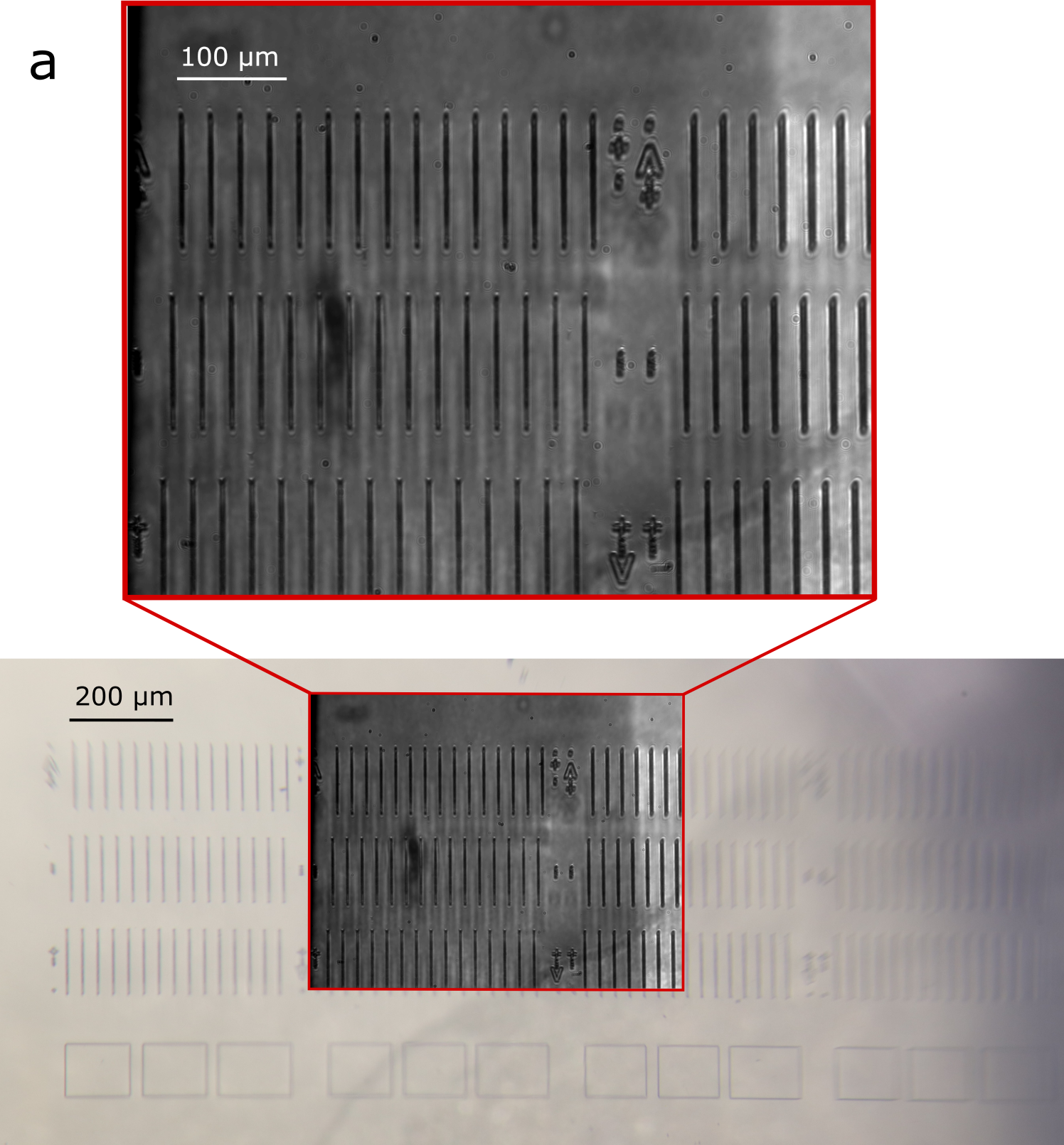
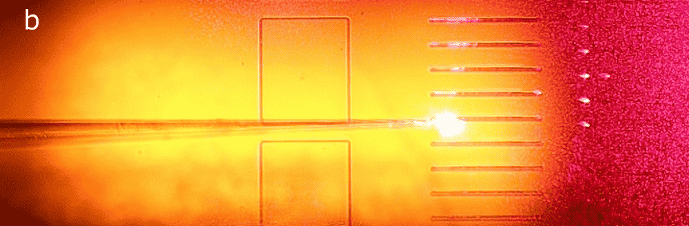
Figure 6: Wide-field and high-magnification images of nanophotonic structures on a diamond chip at ambient and cryogenic conditions.
Field-of-View Dynamics and System Stability
The FOV varies with focal plane shifts induced by vacuum pumping and cooling, as shown by diagonal measurements at the telescope camera. The system maintains a usable FOV of $2.5$ mm, limited by the cryostat's line-of-sight port diameter and optical cutoff for off-axis components.
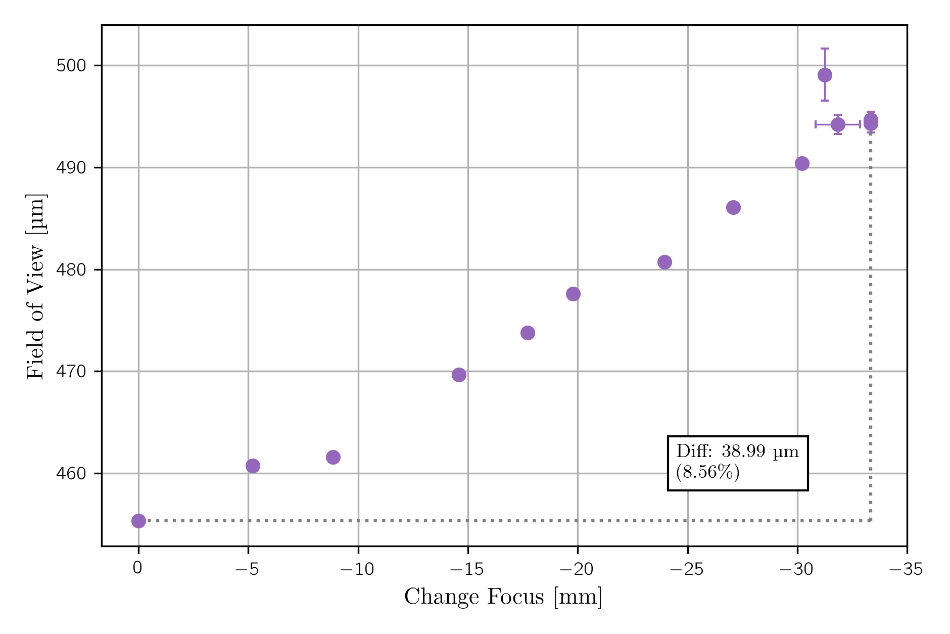
Figure 7: Change of field-of-view measured diagonally due to focus shift from vacuum pumping.
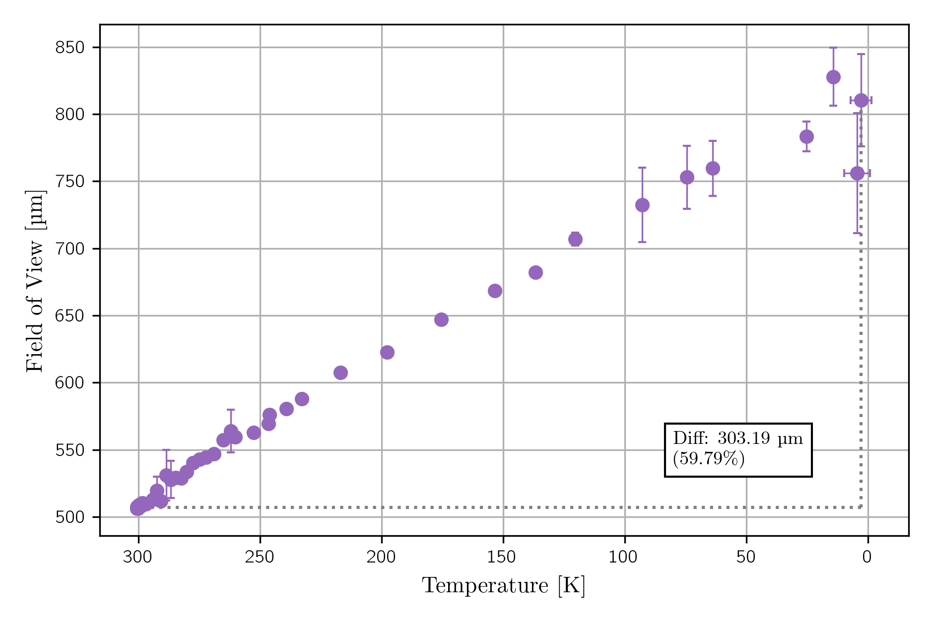
Figure 8: Change of field-of-view measured diagonally due to focus shift from cooling, with higher data collection rate at elevated temperatures.
Magnetic field exposure up to $2$ T along the z-axis and $0.5$ T along x/y-axes did not affect image quality, confirming the suitability of the non-magnetic objective. Vibration from the pulse tube cryocooler introduces axis-dependent resolution degradation, which can be mitigated by mechanical stiffening and synchronization of scanning with the pulse cycle.
Implementation Considerations and Limitations
- Component selection: All optical elements must be rated for low temperature and vacuum operation; achromatic relay lenses are preferred for wavelength-independent performance.
- Thermal anchoring: Gold-plated copper mounts and side rails compensate for differential contraction, ensuring sample-objective stability.
- Focal compensation: Motorized tube lenses and pre-compensation strategies are essential for maintaining focus across thermal cycles.
- Aberration management: Off-axis aberrations and astigmatism require careful alignment and may limit usable FOV for high-resolution imaging.
- Integration with quantum devices: The system supports direct anchoring of temperature-sensitive samples and probe access, enabling in-situ characterization and coupling.
Implications and Future Directions
This imaging system enables high-resolution, low-heat-load visualization of nanophotonic devices at sub-Kelvin temperatures, addressing critical requirements for quantum network node development and optically connected quantum processors. The demonstrated resolution and FOV are sufficient for device characterization, alignment, and fiber coupling procedures. The approach is extensible to other quantum technology platforms requiring stringent environmental control and real-time imaging.
Future developments may include:
- Automated aberration correction: Integration of adaptive optics or computational post-processing to further mitigate off-axis aberrations.
- Enhanced fluorescence imaging: Optimization for confocal fluorescence detection of color centers with higher NA objectives or resonant enhancement.
- Scalable probe station architectures: Modular designs for parallel device testing and integration into quantum network testbeds.
- Real-time feedback for device coupling: Closed-loop control systems leveraging imaging data for automated fiber alignment and coupling optimization.
Conclusion
The reported confocal imaging system in a dilution refrigerator achieves 1.1μm resolution and $2.5$ mm FOV at temperatures down to $10$ mK, with stable performance across thermal cycles and minimal heat load. The architecture supports direct sample anchoring, probe access, and real-time device characterization, facilitating the development of scalable quantum interfaces and network nodes. The technical characterization of focal shifts, resolution dynamics, and system limitations provides a foundation for further optimization and integration into advanced quantum technology platforms.