- The paper introduces a weighted ensemble system that optimally integrates traditional and deep learning models for robust brain tumor classification.
- It employs diverse image preprocessing techniques and feature extraction to capture tumor morphology and enhance predictive accuracy.
- The optimized voting mechanism, validated on Kaggle and Figshare datasets, achieves accuracy up to 99.85%, setting a new benchmark in diagnostic performance.
Optimized Weighted Voting System for Brain Tumor Classification Using MRI Images
Introduction
The paper "Optimized Weighted Voting System for Brain Tumor Classification Using MRI Images" (2603.28357) introduces a weighted ensemble learning framework for MRI-based brain tumor classification. The method synthesizes traditional machine learning algorithms and contemporary deep learning architectures, leveraging complementary data representations and voting-based combination to achieve state-of-the-art predictive performance. This work targets the robust classification of glioma, meningioma, pituitary tumors, and non-tumor cases, addressing the major challenges arising from tumor heterogeneity, morphological variation, and class imbalance.
Methodology
The classification pipeline integrates diverse models: K-Nearest Neighbors (KNN) and Support Vector Machines (SVM) utilizing HOG features and deep architectures including ResNet101, DenseNet121, Xception, a custom CNN-MRI, and ResNet50 applied on edge-enhanced MRIs. The image processing chain applies Balance Contrast Enhancement (BCET), K-means segmentation, and Canny edge detection to maximize intra-tumoral and boundary feature extraction. HOG is employed for shape/texture preservations, while deep models operate on both grayscale and boundary-refined data streams.
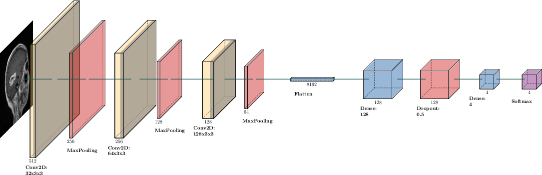
Figure 1: The CNN-MRI architecture used in the experiments.
The motivation for using multiple feature modalities originates from empirical analysis of MR-based descriptive statistics: HOG demonstrates high global-local balance, while edge-based preprocessing (Canny, Sobel) emphasizes anatomical boundaries critical for DL-based discrimination.
All classifiers provide probabilistic class predictions. The final decision employs a weighted voting mechanism, where each classifier's vote is scaled by model-specific validation accuracy. Three weight optimization schemes are explored—uniform, incremental, and automated search—to maximize ensemble performance.
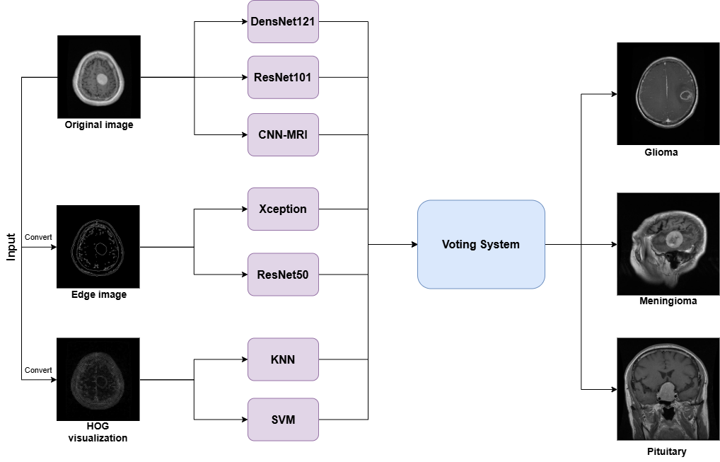
Figure 2: The voting system overview.
Experimental Setup
Experiments utilize the Kaggle and Figshare MRI datasets. The Kaggle set contains 7,023 images with balanced distribution across tumor and non-tumor classes. The Figshare dataset consists of 3,064 images across glioma, meningioma, and pituitary tumors with significant class imbalance. All models are subject to controlled hyperparameter search (AutoKeras for CNN-MRI, Optuna for others), data augmentation (affine transforms, random noise), and standard training schedules. Differences in dataset scale and class balance are explicitly incorporated in model assessment.
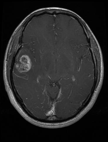
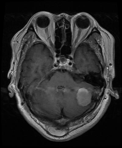
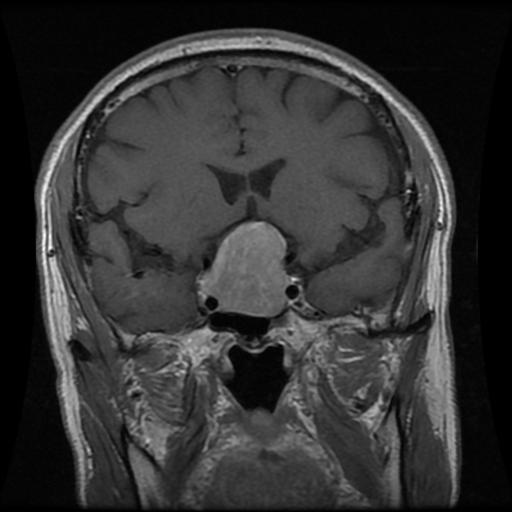

Figure 3: Glioma
Results
DenseNet121 and ResNet101 are the top-performing models, achieving 98–99.6% accuracy on both datasets. Xception and edge-augmented ResNet50 models demonstrate strong spatial feature learning, especially when trained with boundary-focused inputs. KNN and SVM maintain reasonable baselines (>95%) but are regularly outperformed by deep networks.
Weighted Ensemble Analysis
The unweighted (majority) voting achieves 99.13% (Figshare) and 99.54% (Kaggle) accuracy—already exceeding previous hybrid and single-model results. Incremental and highest-weighted schemes show marginal gains or stability on Kaggle data but do not consistently outperform uniform voting under imbalanced Figshare conditions. The automated search for optimal weights further pushes accuracy to 99.46% (Figshare) and 99.85% (Kaggle), indicating that careful weight adaptation—favoring high-performing deep learners—maximizes class-wise F1 and precision, especially for non-tumor splits and pituitary recognition.
Discussion
The ensemble achieves strong numerical results, with 99.85% accuracy on the Kaggle dataset—substantially higher than prior state-of-the-art (typically not exceeding 98.4% in published ensemble/tuning approaches). The performance gain is attributed to the controlled exploitation of classifier diversity and the superior modeling of boundary information via edge detection, as well as optimal parameterized integration of classifier outputs.
The optimization of voting weights elucidates that DenseNet121, ResNet101, and Xception architectures—in conjunction with appropriate image preprocessing—drive decision boundaries for challenging cases (glioma/meningioma confusion) while edge-based augmentations consistently improve sensitivity/specificity for all DL-based routes.
Practically, this paradigm offers a scalable and resilient framework for real-world clinical workflows, where robustness to annotation noise, acquisition variability, and patient heterogeneity is paramount. Theoretically, the empirical dominance of carefully tuned ensembles points to regimes where architectural or feature diversity may supersede architectural scaling, motivating adaptive ensemble strategies in future work.
Implications and Future Directions
- Practical implications: The framework can be readily embedded in diagnostic-support pipelines, particularly as the design is modular—enabling clinician-specific tuning of model composition and voting parameters.
- Theoretical implications: These results reaffirm the importance of feature and architecture diversity in medical image classification, suggesting the limits of single-model generalization in high-variance and imbalanced regimes.
- Future directions involve:
- Dynamic, case-dependent voting (e.g., confidence modeling or meta-learners for weight selection).
- Advanced feature fusion beyond linear voting (attention-based or hierarchical fusion).
- Exploration of additional networks (ViTs, MLP-Mixers), radiomic features, or cross-modality data (CT + MRI integration).
- External validation on larger, multi-institutional, or clinical trial data for robustness verification.
Conclusion
The optimized weighted voting ensemble delineated in this paper demonstrates superior performance in brain tumor MRI classification across both balanced and imbalanced datasets, establishing a new standard for ensemble-based diagnosis-support systems. The analysis confirms that optimal utilization of classifier diversity, boundary-oriented imaging, and architectural advances in DL substantially reduce diagnostic error rates for all major tumor classes. Remaining research gaps concern adaptive weighting for class confusion mitigation and potential integration with interactive, explainable AI systems for radiologists.