- The paper introduces the DL-GIPS framework that integrates deep learning with geometry to synthesize novel-view X-ray projections.
- It utilizes dual encoders for texture and geometry alongside adversarial training to improve image quality over traditional models.
- Experimental results show significant improvements in MAE, RMSE, SSIM, and PSNR, promising reduced radiation exposure in clinical diagnostics.
Novel-view X-ray Projection Synthesis through Geometry-Integrated Deep Learning
The paper "Novel-view X-ray Projection Synthesis through Geometry-Integrated Deep Learning" introduces the DL-GIPS framework. This innovative framework aims to synthesize novel-view X-ray projections by leveraging deep learning techniques integrated with geometric transformations. The objective is to address the limitations posed by traditional multi-angle X-ray imaging, such as increased radiation exposure and complexity in clinical workflows.
DL-GIPS Framework
The DL-GIPS framework comprises four primary components: feature extraction, geometric transformation, image generation, and image discriminator. The process begins with feature extraction from input X-ray projections using two distinct encoders for geometry and texture. These features are integral to ensuring that the synthesized images accurately reflect the new viewpoint while preserving anatomical integrity.
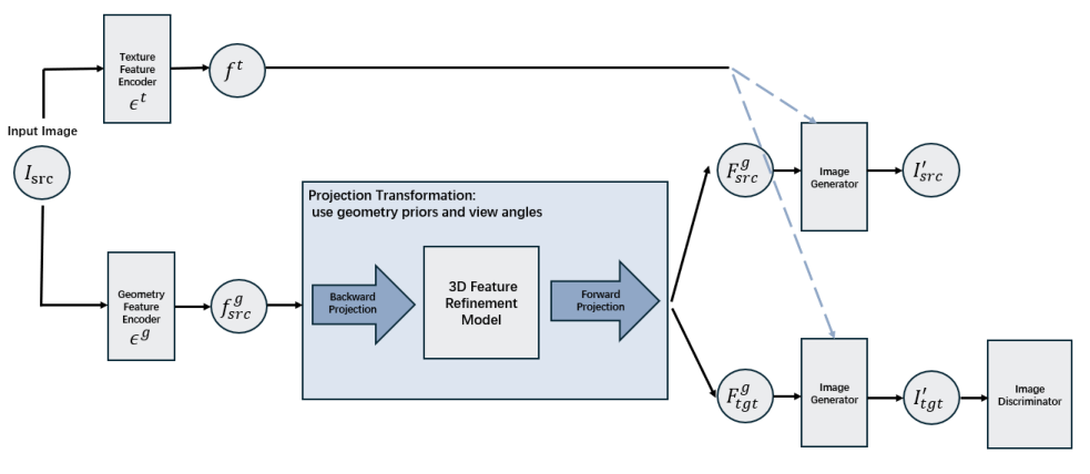
Figure 1: Illustration of the DL-GIPS. The pipeline contains texture and geometry feature encoders, projection transformation, and image generator.
Geometric transformation involves repositioning the extracted geometry features in a 3D space, which are subsequently projected onto the target image plane. This transformation critically relies on cone-beam geometry and necessitates a feature refinement model to address sparsity in input projections.
Image generation synthesizes the new view by integrating the transformed geometry features with texture information. An adversarial training technique is employed, incorporating a multi-scale image discriminator to enhance realism and ensure fidelity to original X-ray projections.
Experimental Evaluation
Two experimental configurations were utilized to validate DL-GIPS: one-to-one and multi-to-multi view synthesis. In the one-to-one scenario, the model synthesized either an anteroposterior (AP) projection from a lateral (LT) projection, or vice versa.
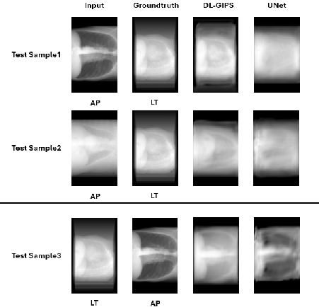
Figure 2: Illustration of one-to-one view synthesis experiments.
In the multi-to-multi configuration, DL-GIPS generated intermediary angle projections (e.g., 30° and 60°) from standard AP (0°) and LT (90°) perspectives.
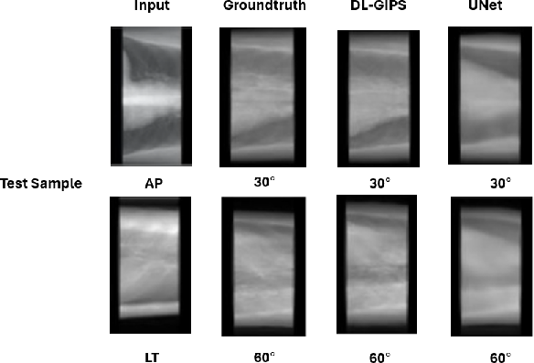
Figure 3: Illustration of multi-to-multi view synthesis experiments.
Quantitative assessments indicate that DL-GIPS substantially enhances synthesized image quality over UNet, particularly in metrics such as mean absolute error (MAE), root mean square error (RMSE), SSIM, and PSNR. For instance, in AP to LT transformation, DL-GIPS achieved an MAE of 0.052 compared to UNet's 0.078. Similarly, PSNR improvements are noted, thereby underscoring the model's capacity to produce superior X-ray prototypes.
Discussions and Implications
The DL-GIPS framework offers significant advancements for X-ray imaging, particularly in scenarios that prioritize reduced radiation exposure—such as pediatric and prenatal diagnostics. By reducing the requisite number of X-ray exposures, this research has implications for enhancing clinical practices while preserving diagnostic accuracy.
However, computational demand remains a pertinent challenge. The geometry transformations necessitate substantial processing power, which currently results in longer inference times compared to conventional models like UNet. Addressing these technological constraints is a priority for future research.
Furthermore, transitioning from simulated datasets to real clinical data is essential for further validation. Future explorations could incorporate "Deep DRR" methodologies for more lifelike X-ray projections, potentially enhancing model generalization and applicability.
Conclusion
Overall, the DL-GIPS model presents a notable enhancement for X-ray imaging by enabling efficient novel view synthesis through a deep learning-geometric integration approach. These advancements suggest meaningful improvements in clinical imaging, offering pathways to minimize radiation exposure while optimizing workflow efficiencies in medical environments. Further developments focusing on computational efficiency and real-world testing will be critical for broader adoption.