- The paper proposes ProstAttention-Net, a deep learning model that integrates a dual-branch CNN with attention mechanisms to segment prostate glands and classify lesions by Gleason score.
- It employs combined cross-entropy and Dice losses with multisource MRI data, achieving sensitivities around 69% and improved performance in the peripheral zone.
- The enhanced segmentation and lesion grading outperform traditional models, indicating strong potential for clinical applications and active surveillance improvements.
Summary of "ProstAttention-Net: A deep attention model for prostate cancer segmentation by aggressiveness in MRI scans"
Introduction
Prostate cancer (PCa) remains one of the most prevalent cancers among men worldwide. Although multiparametric MRI (mp-MRI) has proven effective in detecting PCa, it is challenging to characterize the aggressiveness of prostate lesions using mp-MRI, necessitating biopsies for Gleason scoring. This paper introduces ProstAttention-Net, a deep learning model that segments prostate glands and lesions, grading them by Gleason score. Utilizing an end-to-end multi-class approach, the model incorporates an attention mechanism to integrate anatomical priors, enhancing lesion detection precision.
Methodology
ProstAttention-Net employs a convolutional neural network architecture inspired by U-Net, augmented with attention mechanisms. The network is designed with dual branches: one for prostate segmentation and the other for lesion detection. The segmented prostate gland serves as an attention guide for lesion classification. The model's training involves a heterogeneous series of 219 MRI exams, including data from multiple scanners, enhancing its generalization potential.
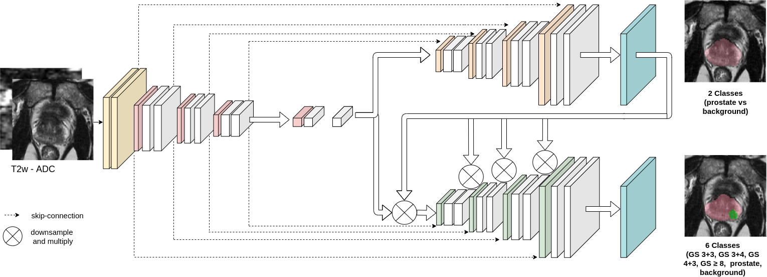
Figure 1: The ProstAttention-Net architecture, illustrating the dual-branch configuration for prostate and lesion segmentation.
The global loss function used combines cross-entropy and Dice losses, weighted to balance class-specific imbalances. This strategic loss formulation aims to optimize both prostate and lesion segmentation accuracy. Hyperparameters are fine-tuned using randomized grid search, and the final model undergoes a validation process with 5-fold cross-validation.
Results
ProstAttention-Net achieved a sensitivity of 69.0% ± 14.5% at 2.9 false positives per patient for clinically significant lesions (GS > 6) detection. When examining the peripheral zone exclusively, the sensitivity improved to 70.8% ± 14.4% at 1.5 false positives per patient. The model demonstrated superior lesion-wise Cohen's kappa coefficients (κ=0.418±0.138), outperforming existing models for automatic Gleason score grading.
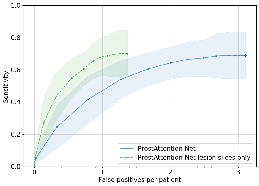
Figure 2: FROC curve demonstrating sensitivity improvements with the inclusion of attention mechanisms.
A comparative analysis against state-of-the-art models, such as U-Net, DeepLabv3+, and Attention U-Net, revealed that ProstAttention-Net offers improved segmentation performance. Notably, the implementation of attention mechanisms is shown to outperform traditional methods, enhancing lesion detection and Gleason score characterization.
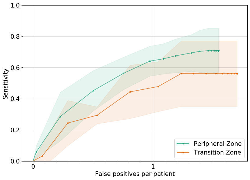
Figure 3: FROC analysis indicating detection sensitivity variations across different prostate zones.
Discussion
The integration of attention mechanisms into the segmentation process offers substantial benefits, particularly in improving lesion grading accuracy. Multisource training data enabled the model to generalize across varied MRI data inputs, although domain adaptation remained a challenge, as evidenced by performance variations on the PROSTATEx-2 dataset.
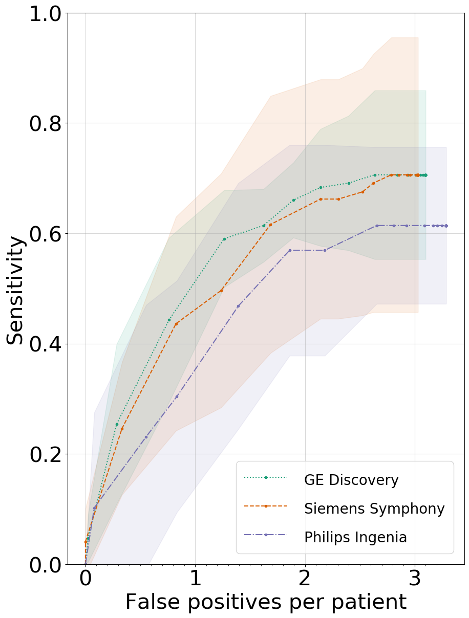
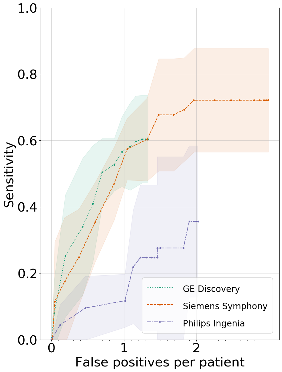
Figure 4: Impact of multisource learning on the FROC sensitivity curves, highlighting generalization capabilities.
These findings validate the model's robustness, although further training data and domain-specific adaptation could enhance performance further. Future research directions may involve integrating additional MRI modalities and exploring weakly-supervised learning approaches to leverage less richly annotated datasets.
Conclusion
ProstAttention-Net establishes itself as a promising tool for PCa segmentation and grading, outperforming traditional architectures in detecting and characterizing PCa. It leverages attention mechanisms to improve anatomical segmentation accuracy, offering novel insights into CAD systems' potential in clinical radiology. The results suggest substantial future utility in clinical settings, particularly in refining active surveillance strategies for lower-grade lesions.
This study marks a crucial step forward in computer-aided diagnostic technology, reflecting the evolving capabilities of deep learning applications in medical imaging.