- The paper demonstrates a novel method for imaging mmWave fields via calibrated Rydberg-state fluorescence and Autler-Townes splitting.
- It employs a three-photon ladder excitation in 87Rb and a steady-state GKSL master equation to extract absolute electric-field amplitudes even in the presence of Doppler broadening.
- The technique enables high-contrast, spatially resolved mmWave field mapping and engineered interference patterns using structured dielectric reflectors.
Calibrated Electric-Field Imaging with Rydberg-State Fluorescence and Autler-Townes Splitting
Introduction and Conceptual Framework
The paper "Calibrated electric-field imaging with Rydberg-state fluorescence and Autler-Townes splitting" (2604.19311) presents a spatially resolved approach for imaging millimeter-wave (mmWave) electric fields inside a warm atomic vapor cell. This technique hinges on Rydberg-state fluorescence, utilizing a multi-photon ladder excitation scheme with a specific decay channel that remains dark except when the mmWave field drives an otherwise forbidden transition. This achieves high-contrast imaging with near-zero background, substantially enhancing sensitivity and spatial discriminability compared to probe-transmission-based methods.
Absolute calibration of local electric fields is realized by reconstructing Autler-Townes (AT) splittings of the Rydberg resonance across the imaging volume. A steady-state solution to the Gorini-Kossakowski-Sudarshan-Lindblad (GKSL) master equation enables robust field extraction, particularly in regimes where spectral features are unresolved due to broadening or Doppler effects. The methodology is further extended to visualize mmWave-standing wave interference patterns and engineer field distributions using structured dielectric reflectors.
Experimental Implementation and Imaging Protocol
The experimental apparatus involves a three-photon ladder excitation in 87Rb: probe, coupling, and Rydberg lasers sequentially access 52S1/2→52P3/2→52D5/2→322F7/2. A mmWave (131 GHz) field drives transitions to 342D5/2, whose decay results in a strong 480 nm fluorescence, detectable with a CCD and bandpass filtering.
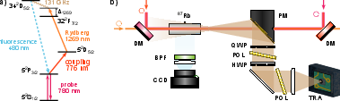
Figure 1: Level diagram and experimental layout including laser paths, polarization control, and mmWave focusing.
The mmWave field establishes a standing-wave pattern, resulting from incident and reflected waves within the vapor cell. Imaging is accomplished by stacking fluorescence images recorded while scanning the Rydberg laser detuning, generating a spatially resolved fluorescence map as a function of laser detuning and position.
To correct optical distortions, an image of a millimeter grid is used to calibrate pixel positions to physical distances. This enables accurate reconstruction of the spatial electric-field profile along the cell, translated from pixel coordinates to millimeters.
Quantitative Analysis and Calibration
Extraction of field amplitudes relies on resolving Autler-Townes splitting, directly proportional to the mmWave Rabi frequency, through fits based on solutions to the GKSL equation rather than simplistic Gaussian doublet separation. This is crucial in room-temperature vapor environments with significant Doppler broadening and other system complexities where the resonance condition is not rigorously satisfied. The fluorescence intensity, assumed proportional to the upper Rydberg population, is fit globally, using manually determined parameters at a representative point and applying the solution to all spatial positions.
Verification is performed using controlled attenuation of the mmWave field via polarization elements (half-wave plate and polarizer), confirming the proportionality between measured Rabi frequencies and expected attenuation, as demonstrated through parity plots.
Engineering Local Field Distributions and Device Applications
The paper explores manipulation of standing wave patterns by integrating a 3D-printed High Impact Polystyrene (HIPS) Bragg reflector. HIPS exhibits low absorption and stable birefringence in the mmWave/THz regime, enabling precise control of reflected amplitude and phase.
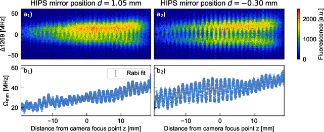
Figure 2: Fluorescence scans for HIPS Bragg reflector in positions minimizing/maximizing fringe visibility, showing normalized intensity and corresponding fitted Rabi frequencies.
By tuning the reflector position, local suppression or enhancement of the standing wave is realized, as visualized with direct overlays of electric-field measurements.
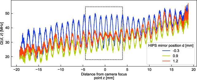
Figure 3: Overlay of E-field measurements for various HIPS mirror positions, demonstrating localized suppression of the electric field.
While perfect suppression was not experimentally achieved, the authors suggest potential for in-situ field measurements and engineering of mmWave-optical interfaces with flexible, scalable dielectric structures.
- The imaging protocol demonstrates spatial resolution well below the mmWave wavelength, set primarily by the beam diameter and camera pixel calibration.
- Field detection sensitivity is governed by the fluorescence channel choice; SNR is vastly improved due to negligible background.
- Absolute calibration via AT splitting allows robust extraction of local electric-field amplitudes even when spectral resolution is limited.
- The methodology offers a minimum detectable field limited by the atomic population and camera sensitivity, with effective suppression/enhancement of field fringes controlled by the HIPS reflector.
Implications, Extensions, and Future Directions
Practically, this approach provides a minimally invasive and calibrated diagnostic for mmWave and THz field mapping in atomic vapor cells, with utility for atomic receiver and sensor calibration, quantum device characterization, and engineered field distribution prototyping.
- Theoretical implications include improved modeling of open quantum systems in field imaging, leveraging advanced master equation solutions for spatially resolved quantitative diagnostics.
- Device applications span high-resolution mmWave imaging, field distribution engineering in hybrid quantum systems, and potential extension to 2D or 3D field mapping using advanced excitation schemes.
- Future developments are proposed: employing alternative fluorescence channels with shorter radiative lifetimes for faster imaging, or light-sheet excitation for two-dimensional mapping.
This method can be integrated with new quantum technologies leveraging Rydberg atoms for electromagnetic field sensing [adams_rydberg_2020], providing a SI-traceable route for mmWave and THz calibration [chen_terahertz_2022], and facilitating the design of miniaturized, print-on-demand components such as reflectors and waveplates [freitas_optical_2025].
Conclusion
This study establishes a self-calibrating, spatially resolved electric-field imaging technique leveraging Rydberg-state fluorescence and Autler-Townes splitting in warm vapor. Absolute calibration, low-background high-contrast imaging, and robust field extraction via master equation fitting distinguish this approach from earlier probe- and transmission-based methods. By visualizing and engineering complex field distributions—including local suppression via printable dielectrics—the methodology offers new capabilities in mmWave-optical interface diagnostics and field sensing. Ongoing and future work may yield advanced imaging geometries, bandwidth/sensitivity optimization, and integration with quantum devices requiring precise electromagnetic control (2604.19311).